7 Easy Facts About Lumbar spine - Radiology Reference Article - Radiopaedia.org Shown


Virtual Spine: lumbar anatomy, 3D model, vertebra, flexion, range of motion, sonoanatomy, ultrasound, regional anesthesia
Lumbar Spinal Cord Injury: What to Expect and How to Manage Can Be Fun For Everyone
Together, they boost the security of the spine and roots. The dura is the most shallow but durable layer. The pia and arachnoid, together termed the leptomeninges, are frail. back pain , roots, and nerve rootlets are closely invested by the pia. The dura and arachnoid together form a loose sheath (called dural/thecal sac) around these structures, separated from the canal walls by the epidural space.
The external surface area is rough and blends with loose connective tissue in the epidural space. The internal surface, facing into the subdural area, is smooth and covered by a layer of mesothelium. Inferiorly, the dural sac ends at the sacral canal, generally at S2-S3 (often S1). The dura continues caudally as a fibrous thread called the filum terminale externum or coccygeal ligament, which mixes with the PLL over the coccyx.

Lumbar Spine Injury L1-L5 - Spinal Cord
Top Guidelines Of Clinical Anatomy of the Lumbosacral Spine - SpringerLink
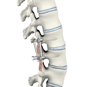
Connective tissue slips in the foramen anchor the dural sleeves so that they can secure the back nerve roots from being stretched during L-spine movements. In addition to these tetherings, the dura is attached in places to the PLL. Epidural space The epidural (peridural/extradural) area ends inferiorly at the sacral hiatus, where it is sealed by the posterior sacrococcygeal ligaments.
The whole area is occupied by loose connective tissue with variable fat material, offering cushioning around the dural sac and spine and functioning as a type to hold the thin internal vertebral plexus of veins open. The vertebral venous plexus is embedded in the epidural loose connective tissue, in some cases transmitting big quantities of blood.
The Best Guide To Magnetic Resonance Imaging of the Lumbar Spine in People
A layer of mesothelium covers all leptomeningeal surface areas bathed by cerebrospinal fluid (CSF). The arachnoid mater lines the whole dural sac and extends into the dural sleeves. It likewise sends trabeculae throughout the subarachnoid space to the pia, assisting in CSF mixing. Along the posterior midline, the trabeculae kind a distinct subarachnoid septum.
The pia mater supplies support for the vasculature and nerves in the subarachnoid space. It adheres intimately to the spine. The pia forms a separate sheath for each nerve rootlet and root as far laterally as the foramen, mixing with the epineurium. Caudally, the pia continues as the thin filum terminale internum.